Clinical Advances - Fall 2020
Clinical Advances
 Call the Fish Doctor
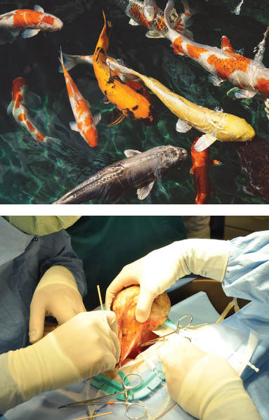
How do you perform surgery on a koi? Very carefully, says Dr. Esteban Soto. His team recently made national news when they removed a softball-sized tumor from Madonna, a 6-year-old koi. Initially, the cause of her distended abdomen was thought to be egg- binding, but a CT scan showed an extensive fluid-filled mass that was pushing on her heart.
During the 90-minute surgery, Madonna was kept anesthetized and alive while out of the water by pumping water containing an anesthetic agent over her gills. It was a delicate balance to keep Madonna from not being anesthetized too deeply or lightly. She remained stable throughout the surgery, and the veterinarians were able to safely remove a 1.1-kilogram gonadal tumor - nearly half her total body weight.
While koi make great pets, Soto is quick to point out that potential owners should understand the amount of work it takes to keep these valuable fish healthy, and to realize that koi can live for more than 100 years.
Beyond health examinations and surgeries on a variety of fish species, Soto also performs water testing, virus testing, and necropsies through the Aquatic Animal Health unit of the Companion Exotic Animal Medicine and Surgery Service. His primary objective is to raise fish health awareness and provide medical services to private owners, hobbyists, breeders, wholesalers, retailers, commercial aquaculture, aquariums, and referring veterinarians, as well as government fish and wildlife conservation agencies. Soto was critical in the Veterinary Emergency Response Team’s rescue of dozens of koi during the 2017 Napa fires. Most were estimated to be between 30–70 years of age.
 Something to Meow About
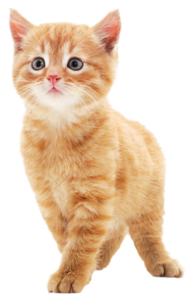
A hospital stay can be stressful for any animal, but particularly for cats who may not be familiar with leaving home. To help minimize their anxiety, and provide a more efficient treatment area, the UC Davis veterinary hospital created a new, all-inclusive feline suite combining a hospitalization ward with examination and treatment space. Veterinarians and technicians laud the suite’s efficiency in helping them provide optimal care for multiple hospitalized cats.
These renovations to the small animal clinic are part of Phase I of the future Veterinary Medical Center, and include six new examination rooms, along with upgrades to the laundry and support facilities, locker rooms and restrooms. Funding for this new feline facility was provided by an anonymous donor who is dedicated to the advancement of cat health.
Space can be a big issue during wildfires or other large community emergencies, when 50–75 cats may need to be hospitalized at the same time. This new facility allows the veterinary team to treat and house the cats in an all-in-one suite, eliminating the need to transport them from a hospitalization ward to an examination room in another part of the hospital.
“This three-room facility will completely transform patient care for more than 5,000 cats we see annually and allow us to do the very best for them,” said Dr. Kate Hopper, director of the Small Animal Clinic.
FELINE SUITE FEATURES
- Expanded hospitalization ward for more than 20 cats
- Large 2-table examination/ treatment area
- Private 1-table examination/ treatment area
- Animal washing area
- Equipment/supplies station
- Sound dampening throughout to decrease stress-inducing noise
- Emergency eye wash station
- Work stations for four technicians and veterinarians
Saving Lives and Livelihoods
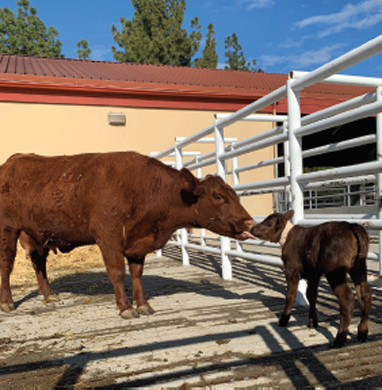 When a sudden illness on the Schuler Ranch started sweeping through a herd of newborn calves, killing one of them, fourth-generation rancher Brian Greathouse immediately contacted the Livestock Medicine and Surgery Service. At two in the morning, the on-call and on-site veterinary team helped the ranch prepare the affected cattle for transport to the hospital.
Thanks to UC Davis’ unique ability to perform a necropsy onsite at the hospital, as well as process samples at our cutting-edge lab facility, clinicians were able to provide a quick diagnosis and initiate an urgent treatment plan for the remaining calves. They were infected with K99 Escherichia coli (“E. coli”), as well as Cryptosporidium parvum (“crypto”). These bacterium and parasites can cause severe diarrhea and be deadly to newborns.
Proper vaccination and herd health are top priorities at the Schuler Ranch, so they quickly commenced a thorough investigation. It was determined that the most likely cause for increased exposure to E. coli or crypto came from geese migrating through and resting in the pastures.
Thankfully, there is a vaccination for K99 E. coli that could be used on the pregnant cows and heifers to help prevent and/or limit impact of the disease in the other calves. The rest of the calving season at the Schuler Ranch was back to “business as normal,” with the remaining cows moved to a different pasture (that the geese had not migrated through) to calve out.
The four hospitalized calves were cared for by the Livestock Medicine and Surgery Service with IV fluid therapy, antibiotics and pain management. All responded well and recovered quickly. The mother cows accompanied the calves to the hospital to continue delivering nutrient-rich milk. Equally important, the mothers avoided mastitis by having their udders emptied daily.
“We’ve brought our animals to UC Davis for more than two decades,” Greathouse said. “In this case, they saved us quite a bit of financial loss. But more importantly, they saved those calves’ lives. We’re not a big outfit, so what everyone at UC Davis did helped us survive a potentially devastating loss to our operation.”
Revolutionizing Orthopedics
When Luna, a one-year-old female Bernese mountain dog, jumped out of a moving car, she broke the head of her right femur. As a champion show dog, Luna’s best chance of recovering a normal gait was to undergo a total hip replacement, so her local veterinarian referred her case to Dr. Denis Marcellin-Little, UC Davis’ renowned orthopedic surgeon. Luna healed well enough to return to the show ring.
When Marcellin-Little joined the school as a professor of orthopedic surgery in 2017, he sought to advance fracture repair research and expand surgical options for complex surgeries like Luna’s total hip replacement.
Collaborating with the Veterinary Orthopedic Research Laboratory and UC Davis biomedical engineers, Marcellin-Little is able to utilize 3D printing and other technology to create custom implants to improve surgical outcomes for patients like Luna, as well as revolutionize fracture repair in smaller dogs.
Marcellin-Little works with computer numerical control (CNC) milling machines to fabricate a piece of metal or other material (such as polymer) into a specific object, based on coded programmed instructions – similar to the technology behind 3D printing. Marcellin-Little can utilize the CNC technology to create specific plates that will ultimately be used to repair a dog’s broken radius in instances where commercially available plates will not work. This plate research could drastically change the way orthopedic surgeons handle fractures that are not healing and may lead to amputations.
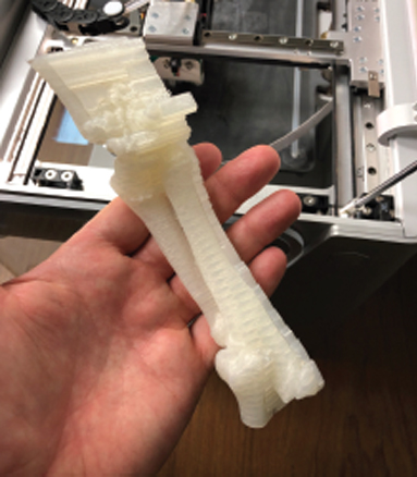
Similarly, Marcellin-Little uses 3D printing to open new doors into surgical procedures where commercial hip replacement kits might not be the best fit for a dog. Utilizing exact anatomical measurements from a CT scan, Marcellin-Little works with a biomedical manufacturing company to craft a custom, 3D-printed titanium implant. These state- of-the-art implants can even be coated with silver to reduce chances of bacterial growth and stave off potential infections. While these custom hip replacements are being produced, Marcellin-Little and his surgery team create exact replicas of a dog’s bones with in-house 3D printers to practice the procedure prior to surgery.