Clinical Updates
Expanding Care
State-of-the-art equipment, advanced treatment options, telemedicine and surgical expertise expand the limits of what UC Davis offers to veterinary patients.
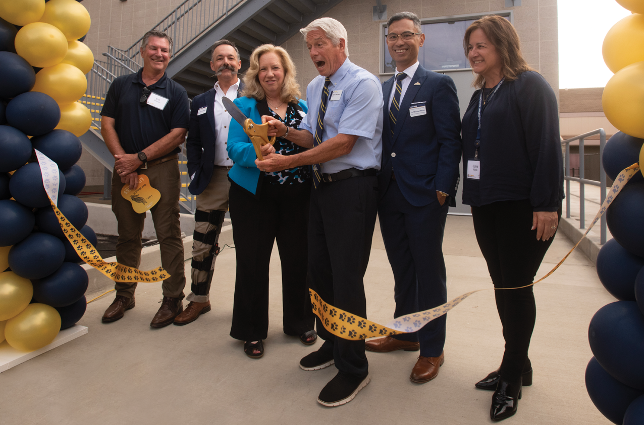
All Species Imaging Center Opens
After years of careful planning and construction, the UC Davis William R. Pritchard Veterinary Medical Teaching Hospital (VMTH) opened the All Species Imaging Center in September. Having a central hub for the hospital’s advanced diagnostic imaging is the realization of leadership vision and philanthropic endeavors nearly a decade in the making. The project was made possible thanks to the generosity of many, as the entire building and two of the new scanners were donor funded.
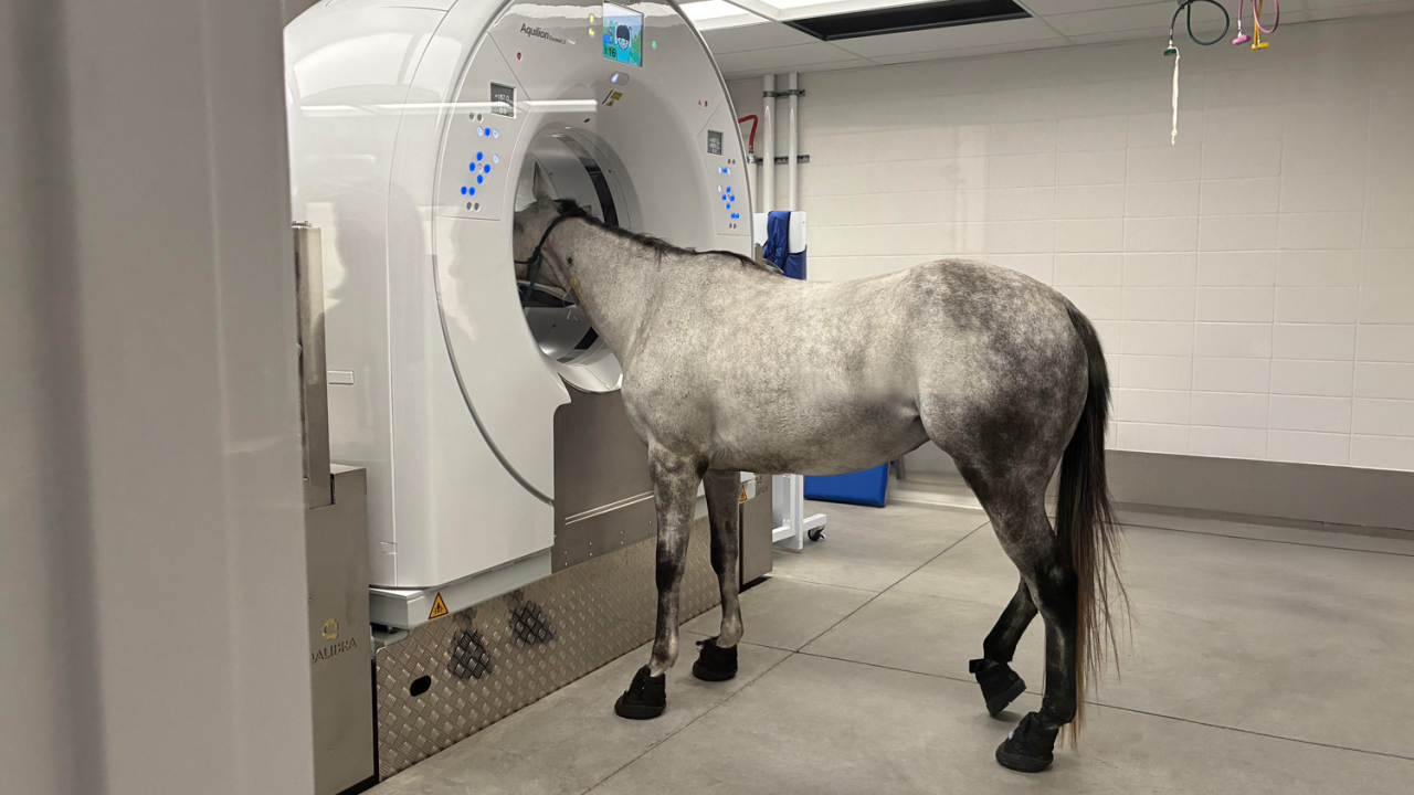
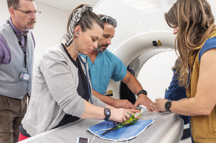
The imaging center is adjacent to the VMTH and includes four diagnostic imaging suites, two control rooms, a radiologist’s image interpretation room, and office space for technical staff. The four imaging suites are dedicated to: small animal computed tomography (CT); high field magnetic resonance imaging (MRI) for small and large animals; positron emission tomography (PET)/ CT for small and large animals; and a dedicated large bore equine CT, which is revolutionizing how the hospital diagnoses and treats horses.
The new small animal CT scanner and the MRI scanner replaced equipment that had been in use at the VMTH for more than 15 and 20 years respectively. These state-of-the-art machines offer higher image quality and faster image acquisition. They also open the door for advanced clinical applications such as 4D cardiac imaging, functional brain imaging, and novel clinical diagnostic studies. Thanks to these advanced technologies, UC Davis is now able to increase diagnostic imaging caseload and fulfill the demand for the highest quality imaging services in California.
The equine CT scanner allows head and distal limb scans on standing horses that are only under sedation vs full anesthesia. The mobility of the scanner due to its integration onto a complex platform that can be moved both vertically and horizontally, combined with the large bore of the scanner, allows for CT imaging of the stifles, pelvis, and entire vertebral column. This is the first CT scanner/ platform combination of its type to be commissioned in the state.
The center also serves as home to the world’s largest advanced radiology training program. With 12 residents and eight board-certified radiologists, the hospital’s Diagnostic Imaging Service is the premier training ground for veterinarians seeking board certification in imaging.
Stomatitis Clinic Soaring to New Heights
As the Dentistry and Oral Surgery Service’s (DOSS) Stomatitis Clinic commemorates its third anniversary, they also celebrate the continued success of clinical trials to treat the disease, which causes painful inflammation of the mouth and gums.
While UC Davis has been pioneering treatments for feline stomatitis for more than a decade, clinical trials have now expanded to include dogs. Lulu, an 11-yearold miniature Schnauzer, was treated for canine chronic ulcerative stomatitis, which caused her years of severe oral pain. Following extraction of her compromised teeth, Lulu was enrolled in a clinical trial for triple therapy consisting of an antibiotic, a form of vitamin B3, and a medication used to improve blood flow. She showed significant improvement after completion of the 4-month trial and remains in full remission.
While UC Davis has been pioneering treatments for feline stomatitis for more than a decade, clinical trials have now expanded to include dogs.
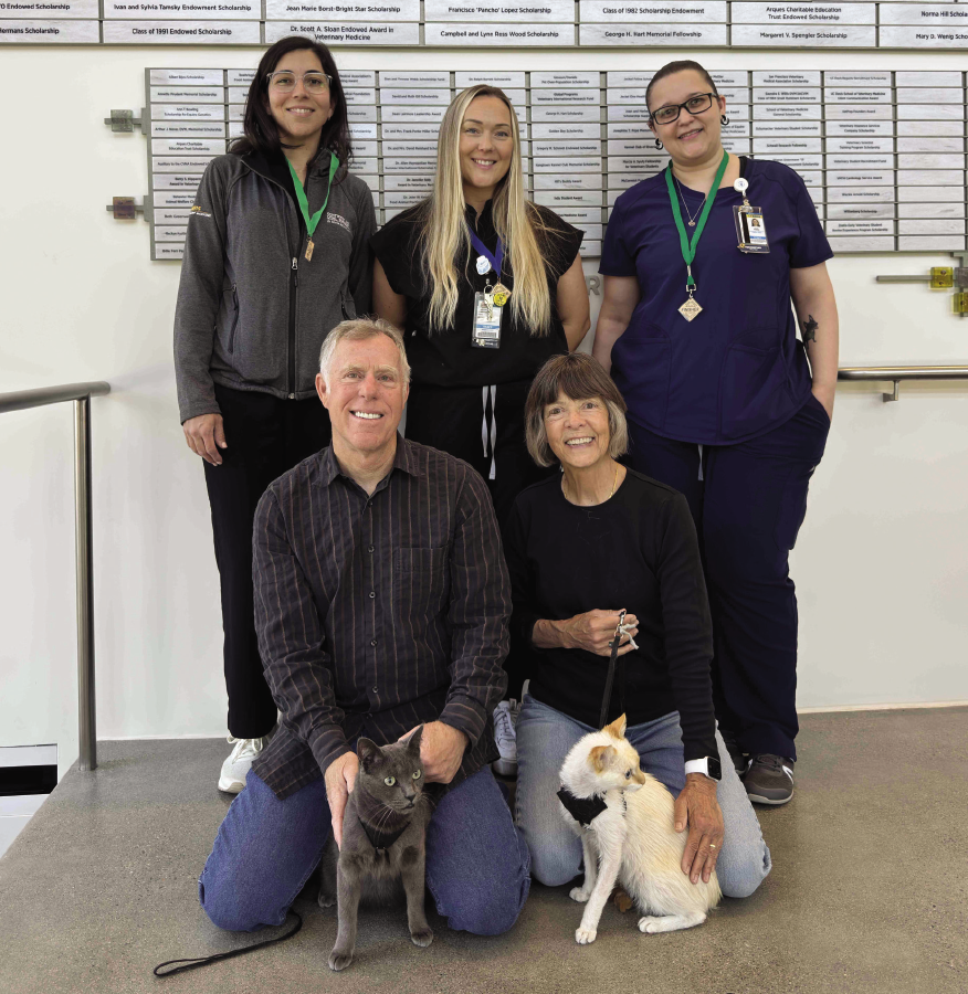
Continuing the clinic’s success of stomatitis research and care in cats, Dr. Maria Soltero-Rivera is studying the further use of stem cells to treat feline chronic gingivostomatitis (FCGS). A previous trial led by Dr. Boaz Arzi showed remission success of 70% when stem cells were administered two months after teeth extraction. Soltero-Rivera just completed a trial that administers the cells at the time of extraction and is now conducting a Gallant-sponsored trial for patients that show resistance to treatment.
Robin, a 2-year-old gray Domestic Shorthair cat, is in remission from FCGS after completing the Gallant trial. Robin’s owners, Steve and Jean Gill, were so impressed with the success that they made two gifts to support Soltero-Rivera’s research and in honor of Robin’s care team.
Due to its capacity caseload, DOSS is only able to dedicate two days per month to the Stomatitis Clinic, but that will double next summer with the creation of an oral medicine fellowship that will add a veterinarian to the team. This unique, 2-year advanced training program will allow a board-certified dentistry specialist to hone their skills in oral medicine and research, while gaining valuable clinical experience treating stomatitis patients.
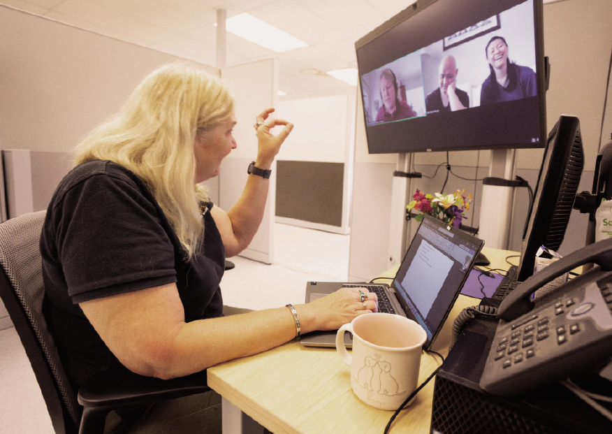
Hospital Offers Telemedicine Appointments
In 2024, a new California law established that new veterinary-client-patientrelationships could be made via telemedicine appointments. This means that initial in-person examinations are no longer necessary to begin seeing new patients. Since the law passed, UC Davis has established telemedicine appointments in four specialties: behavior, fish medicine, nutrition, and orthopedic surgery. More specialty services may be added as this convenience becomes more readily available for our clients.
A telemedicine appointment is a prescheduled one-on-one consultation with a veterinarian via the Zoom communications platform that allows users to connect with video and audio on a smartphone or computer. This allows veterinarians to observe a pet and diagnose a clearly visible health condition such as a rash, laceration or limp.
There are some limitations and legal requirements of telemedicine consultations at UC Davis, most importantly that appointments are only available to California residents. The same standards and laws governing veterinary practice apply to telemedicine and in-person veterinary medical services. However, California state law places certain limits on a veterinarian’s ability to prescribe medications using telemedicine. While antimicrobials (antibiotics, antifungals, etc.) can be prescribed for up to 14 days, controlled drugs (opioids, sedatives, etc.) cannot be prescribed. Veterinarians can only prescribe long-term medications for up to six months and cannot prescribe drugs off-label.
Telemedicine has long proved to be a valuable tool increasing efficiency
of processes in consultation and treatment.
Telemedicine has long proved to be a valuable tool increasing efficiency of processes in consultation and treatment. Prior to the new law, several specialty services at the veterinary hospital were allowed to use telemedicine appointments to pre-screen patients before the initial inperson consultation and to perform recheck evaluations when diagnostic testing and/or hands-on evaluations were not necessary.
During the pandemic, the Behavior Service often utilized telemedicine appointments after initial evaluations were conducted on campus. These virtual “house calls” worked well for clients and veterinarians alike and are now made even more convenient without the initial in-person requirement. Behaviorists comment that seeing pets interact with their owners in their home environments presents a more natural view of their potential behavior issues. Virtual visits also keep animals safe in their home environment, especially beneficial for dogs being treated for anxiety.
While this new application of technology greatly enhances accessibility and availability of specialty care throughout California, it will not replace in-person visits. Unlike human medicine where patients can verbalize their symptoms to their physician, veterinarians rely heavily on the insight they obtain via physical examination. Despite this limitation, telemedicine is bound to have an impact on veterinary medicine, and UC Davis is positioning itself to enhance its reach by harnessing the technology.
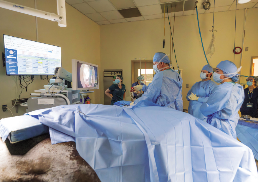
Foraminotomy Surgery Now Available for Horses
An extremely specialized equine surgery performed by only about a dozen veterinarians nationwide is now available at UC Davis. Thanks to the addition of Dr. Carter Judy to faculty, as well as a dedicated large bore equine CT scanner, the veterinary hospital offers foraminotomy surgery for horses with cervical foraminal stenosis—a narrowing of an intervertebral opening where the limb nerve root exits the spinal column.
The condition can be caused by arthritis, bone spurs, disc herniation, or thickened ligaments. It places compression on the nerve resulting in pain and numbness that can develop into lameness and behavior issues such as ataxia, skipping gait, and headshaking.
Diagnosing the condition—which generally occurs near the base of the neck where cervical and thoracic vertebrae meet—requires a large bore equine CT scanner, which was recently installed in the new imaging center. Prior to obtaining that equipment, the veterinary hospital had a traditional CT scanner whose configuration did not allow for imaging beyond a horse’s head and distal limbs.
Foraminotomies were first performed on horses in Europe in 2020. Similar surgeries in humans have existed for several decades. Foraminotomy surgery is performed minimally invasively, using endoscopic instruments to burr the foramen larger, relieving pressure on the compromised nerve root. Many horses show improvement less than 24 hours after surgery.
The hospital is attracting clients from around the nation due to the limited number of surgeons who perform the procedure.
Judy performed the surgery for a year at his previous clinic before recently joining the UC Davis faculty. Even without an equine CT scanner onsite when he first arrived, he was able to perform the surgeries immediately at UC Davis with imaging acquired from other hospitals. But now with the large bore CT scanner, the wait times and travel requirements for horses with this condition are greatly reduced.
Drs. Scott Katzman and Heidi Reesink completed foraminotomy training this year, allowing more appointment opportunities for this unique surgery. The hospital is attracting clients from around the nation due to the limited number of surgeons who perform the procedure.
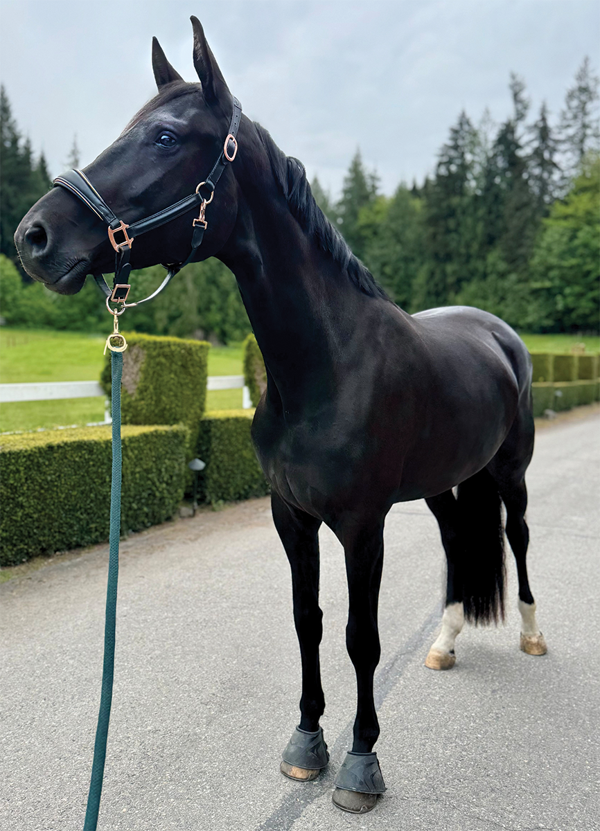
One such horse who had suffered from this condition is Frida, a 4-year-old Oldenburg mare from Washington. Osteoarthritis of multiple facet joints had caused foraminal stenosis, resulting in hind-limb weakness, range-of-motion and coordination problems. Foraminotomy surgery corrected these conditions, and she continues to improve.