Clinical Updates
Advancing Clinical Care
New facilities, equipment, technologies and collaborations push the boundaries of care for our companion animals and wildlife.
Compassionate care has always been the gold standard at UC Davis—whether the patient is a pet hamster, a best-in-show dog or an injured wild hawk. That level of care is enhanced when our veterinary specialists collaborate in expanded clinical facilities where advanced equipment allows them to employ new technologies and surgical approaches.
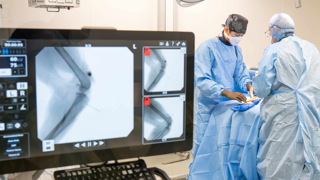
Surgery Center Celebrates First Year
Sambatra, a 4-year-old male ring-tailed lemur housed at the Sacramento Zoo, came to UC Davis for surgery after sustaining a knee injury. He was one of more than 500 patients treated in the first year of the school’s Advanced Veterinary Surgery Center. Repair of Sambatra’s ruptured patella ligament was a perfect fit for the new center’s orthopedic surgery focus.
Built entirely with donor funds, the center allows for increased surgery appointments and is reducing wait times for non-emergent orthopedic cases. Clients and their pets have access to a team of renowned orthopedic veterinary surgeons including: Drs. Po-Yen Chou, Barbro Filliquist, Denis Marcellin-Little, and Amy Kapatkin, who was awarded the 2024 American College of Veterinary Surgeons (ACVS) Founders’ Award for Career Achievement. The award recognizes the service of an ACVS Diplomate distinguished by contributions to the development of surgical techniques and methodology and disseminating knowledge to colleagues, residents, and students.
Our surgical specialists set the bar for innovative treatments.”
—Dr. Mark Stetter
Training under these surgeons is a much sought-after position, as the center is enhancing opportunities for the next generation of specialists through resident and fellowship opportunities. The advanced level of cases seen at the surgery center amplifies UC Davis as the leading house officer program in the country. The surgical residency currently hosts six positions, and one fellow—board-certified surgeon Dr. Ming Lu, who is participating in a joint replacement fellowship.
“Our surgical specialists set the bar for innovative treatments,” said Dr. Mark Stetter, dean of the school. “With the surgery center, we are motivated to grow our capacity to lead a rapidly evolving field to even greater heights.”
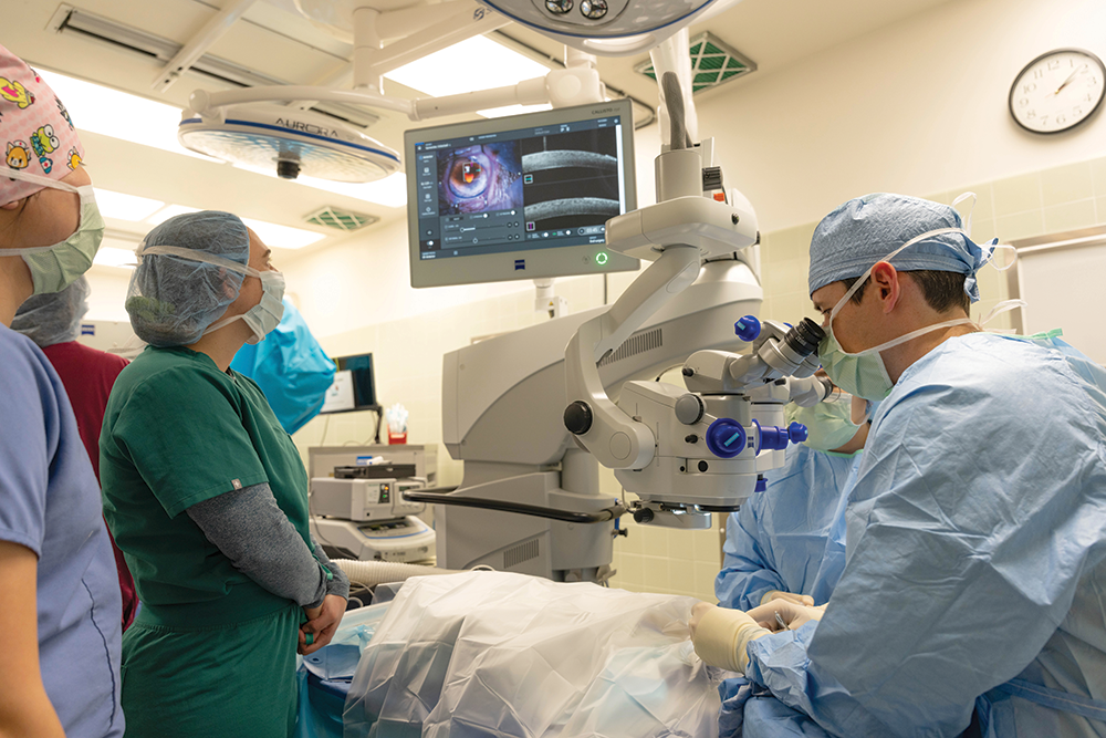
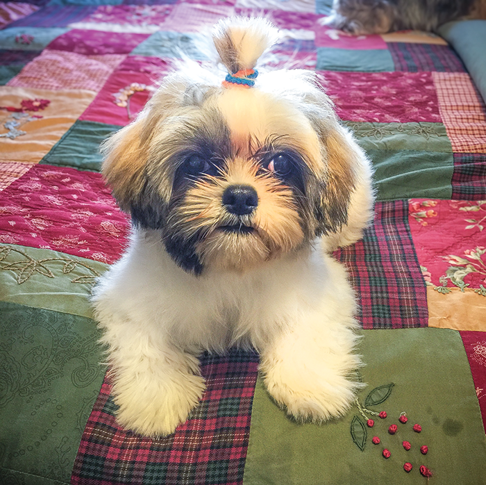
Expanding Vision
When Tashi’s owner Jerry Nordeen noticed changes in her behavior such as confused staring, bumping into things, and the inability to judge distance, he suspected a problem with her eyesight. A visit with the 7-year old Shih Tzu’s veterinary ophthalmologist confirmed a diagnosis of bilateral retinal detachment and the dog was referred to UC Davis for advanced care.
Thanks to a new microscope recently acquired by the Ophthalmology Service, as well as additional surgical equipment funded by a donor, retinal reattachment surgery is now available at UC Davis.
One of the microscope’s many features is a back-of-the-eye viewing system that offers surgeons a clear and uncompromised view of the retina and other anatomical areas at the back of the eye. The new equipment saved Nordeen a trip to Southern California, the next closest place for the surgery. Only a handful of other clinics nationwide have the capability to perform the delicate procedure.
Advanced imaging components in the microscope, including Resight—an automated viewing system for the posterior structures of the eye—and Optical Coherence Tomography (OCT), assists clinicians in visualizing areas beyond the retina. Previously, ophthalmologists could only rely on OCT as a pre-surgery assistance tool; the new microscope allows for near-histologic detail images (microscopic images used to help diagnose disease) to be taken of the eye during surgery.
The new microscope is also proving valuable in cataract surgeries and foreign object removals, which are now performed more efficiently with the use of OCT during surgery. OCT images can be displayed in double-depth resolution in the surgeon’s view to better visualize small details like the location of a foreign object relative to a vital part of the eye.
Thanks to this advanced imaging, many patients like Tashi enjoy renewed sight and improved quality of life.
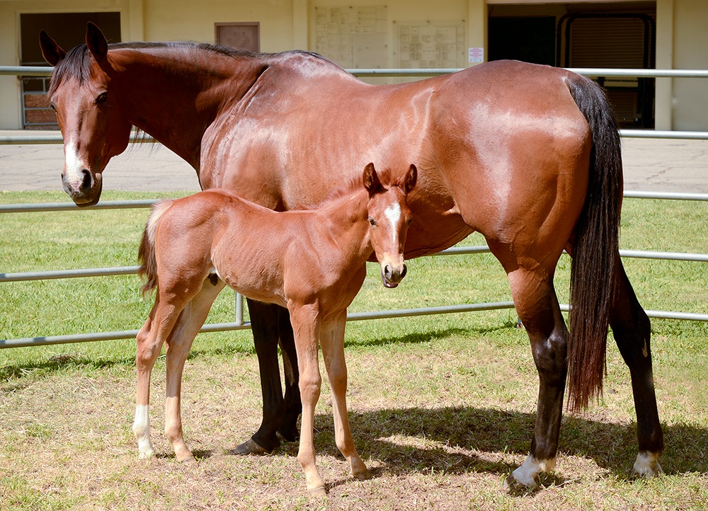
Making Strides in Equine Reproduction Advancements
UC Davis’ long-standing relationship with the horse industry in California and around the world has led to a wide range of advances in equine reproduction. Because of this, the Equine Reproduction Service has experienced significant growth over the past few years in terms of staffing and technology. The service team has grown to seven veterinarians (three faculty, two staff, and two residents) and increased their caseload by 10% last year.
UC Davis has pioneered the advancement of intracytoplasmic sperm injection (ICSI)—the current IVF technology for producing foals.
Working with the school’s Veterinary Assisted Reproductive Technology Laboratory (VetART), the service is expanding the limits of reproduction technology. The VetART Laboratory recently became the first in the world to produce equine embryos through in vitro fertilization (IVF) using frozen sperm. While a foal has not been born yet through this process, this milestone in equine assisted reproductive technology has the potential for broader clinical applications, as well as valuable opportunities to study fertilization and embryo development.
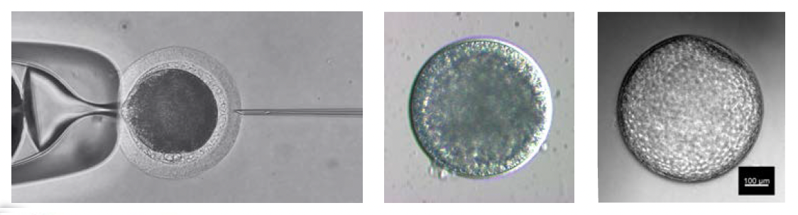
UC Davis has pioneered the advancement of intracytoplasmic sperm injection (ICSI)—the current IVF technology for producing foals. ICSI involves injecting a single sperm into an egg collected from a mare. The embryo then develops in the lab for a week before being implanted in the mare. This is a growing field, and in 2024 alone, UC Davis produced 560 embryos through ICSI for horse owners throughout the United States.
The progress VetART and the Equine Reproduction Service are making together is exciting news for horse owners who may be struggling to breed their low fertility mares and stallions.
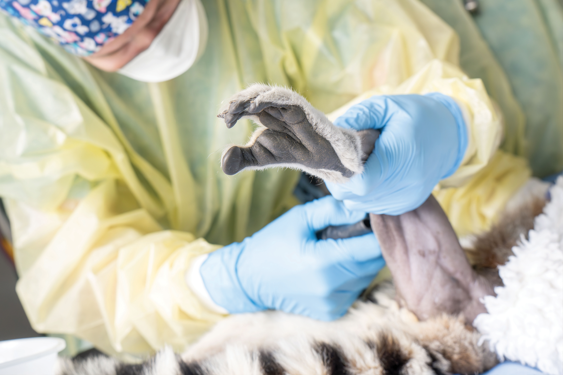
Exotics Care Covers Many Species
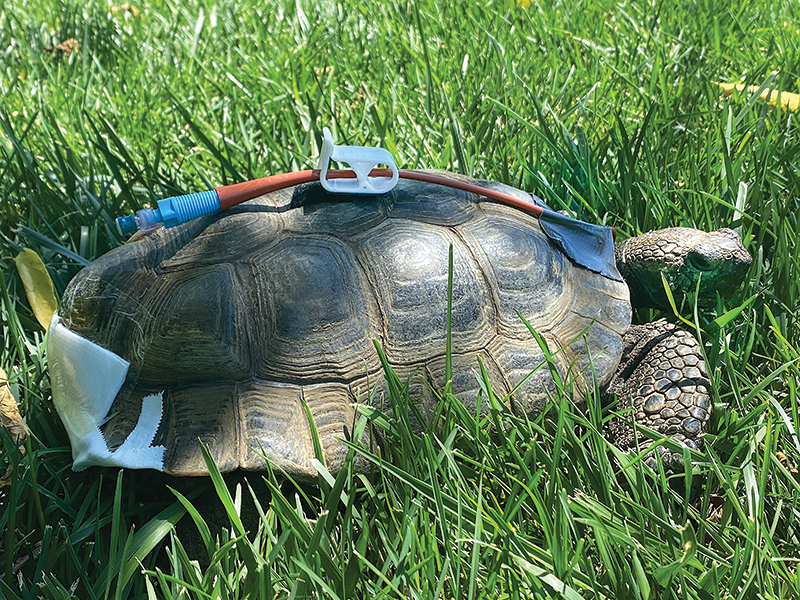
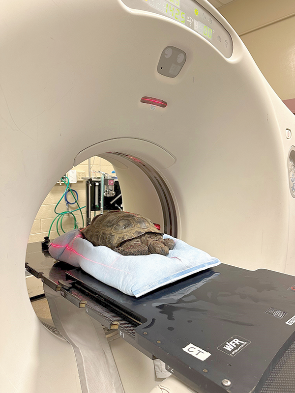
Teetle, an 80-year-old male California desert tortoise, was brought to UC Davis for treatment of a large mass—later determined to be an osteosarcoma—growing beneath his shell. Exotic pet specialists and soft tissue surgeons collaborated on a novel procedure to access the cancerous tumor. They had to remove a portion of his upper shell to minimize disruption of the supportive muscles of his lower back, vertebral column, and hind limbs.
Teetle is just one of thousands of patients the Companion Exotic Animal Medicine and Surgery Service treats every year. The service routinely sees birds, rabbits, ferrets, guinea pigs, chinchillas, rats, hamsters, snakes, lizards, turtles, and many other exotic animals in its thriving practice. Their patients are often the talk of the hospital, especially when an 80-year-old tortoise arrives. The service’s five faculty veterinarians and one staff veterinarian divide the caseload among their extensive experience and certifications in different areas of exotic animal care.
Dr. David Guzman is board certified in both the American (ACZM) and European Colleges of Zoological Medicine (ECZM) with emphases in avian and small mammal medicine. Dr. Hugues Beaufrere is triple board certified by ACZM, ECZM (avian), and the American Board of Veterinary Practitioners (ABVP) in avian practice. Dr. Michelle Hawkins is also ABVP board certified in avian practice and is the director of the school’s California Raptor Center, where birds of prey are rehabilitated for release. Dr. Krista Keller is board certified by ACZM.
The team even has a fish specialist, Dr. Esteban Soto, who is a certified aquatic veterinarian with board certifications by the American College of Veterinary Microbiologists (ACVM) and his newly acquired board certification by ABVP with a specialization in fish practice. Rounding out the team is staff veterinarian Dr. Nicole Mikoni, who recently completed her residency in zoological medicine at UC Davis and is pursuing board certification.
The service is happy to report that Teetle is recovering well at home, and his owners say that he has more energy than ever since his surgery.